Ultrasound Scans: How Do They Work?
Ultrasound imaging, also known as sonography, is a medical imaging technique that uses high-frequency sound waves to produce images of internal body structures. It is a non-invasive and painless procedure that has become an essential diagnostic tool in modern medicine. Visit Beautiful Beginnings Ultrasound Clinic today to get specialized and comprehensive care. For more information, contact us today or book an appointment online. We are conveniently located at 180 Ave at the Cmns Suite 9, Shrewsbury, NJ 07702.


Table of Contents:
What happens during an ultrasound?
Are ultrasounds safe?
When should I know the results of my ultrasound?
What conditions can be detected by an ultrasound?
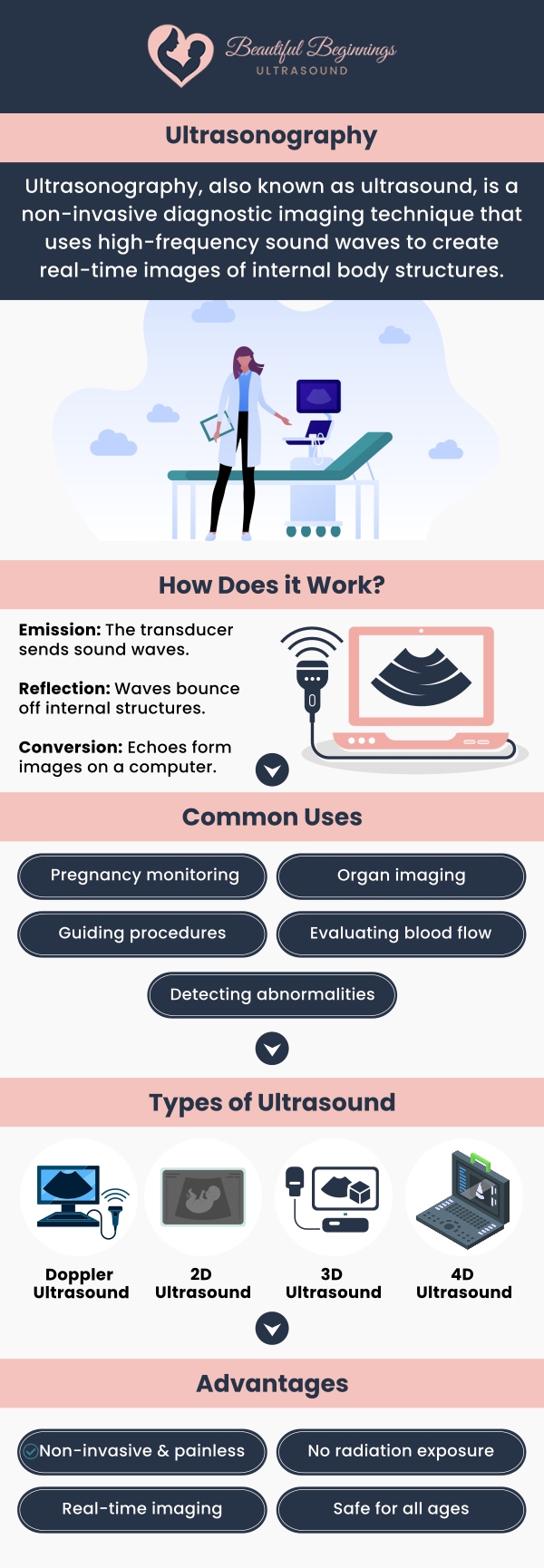
The first step in the process of ultrasound imaging is to prepare the patient for the procedure. This involves removing any clothing or jewelry that might interfere with the imaging process and positioning the patient in a way that allows for optimal visualization of the area being imaged. Once the patient is positioned correctly, a gel is applied to the skin to help transmit the sound waves from the ultrasound equipment. The ultrasound equipment consists of a transducer, which emits high-frequency sound waves, and a computer, which processes the echoes that bounce back from the internal body structures. The transducer is placed on the skin and moved around the area being imaged to obtain different views.
There are several types of ultrasound imaging techniques, each with its own advantages and limitations. 2D ultrasound imaging is the most common type and produces two-dimensional images of the internal body structures. 3D and 4D ultrasound imaging, on the other hand, provide three-dimensional images that offer a more detailed view of the internal structures. Doppler ultrasound imaging is another type of ultrasound that uses sound waves to measure the speed and direction of blood flow in the body. This technique is commonly used in cardiology and vascular imaging.
Ultrasound imaging has a wide range of applications in different medical fields. In obstetrics and gynecology, an ultrasound is used to monitor fetal development and detect any abnormalities in the reproductive system. In cardiology and vascular imaging, ultrasounds are used to detect and monitor heart and blood vessel diseases. In abdominal and gastrointestinal imaging, an ultrasound is used to diagnose and monitor diseases of the liver, pancreas, and other organs in the digestive system. Ultrasound imaging is also used in other medical fields, such as urology, neurology, and musculoskeletal imaging.
Ultrasounds performed by a doctor are safe for you and your baby. Because an ultrasound uses sound waves rather than radiation, it is safer than X-rays. The supplier has been using ultrasounds for more than 30 years and has not identified any dangerous risks.
An ultrasound is a non-invasive medical imaging technique that uses high-frequency sound waves to create images of internal organs and tissues. Although it is most associated with pregnancy scans, ultrasounds have a wide range of applications in both medical and non-medical fields.
Most ultrasound exams take 30 minutes to an hour to complete. In most cases, the results will be provided to you immediately after the exam is completed, but the actual images may need to be analyzed by a radiologist.
Ultrasounds are widely used in medicine for diagnostic imaging of internal organs and tissues. It is particularly useful for identifying abnormalities in the liver, kidneys, and reproductive organs. An ultrasound is also commonly used to monitor fetal development during pregnancy, as it can detect problems such as an ectopic pregnancy, multiple pregnancies, and fetal abnormalities. In addition, ultrasounds can be used to evaluate blood flow and circulation, particularly in the heart and blood vessels.
Ultrasounds have a variety of nonmedical applications, including industrial inspections of materials and structures. It is commonly used to detect flaws and defects in machinery and equipment, as well as to clean and process surfaces and materials. For example, ultrasonic cleaning is used to remove dirt and contaminants from delicate or complex parts, such as electronic components and medical devices. Ultrasonic testing is also used in the aerospace and automotive industries to inspect materials for defects and damage.
Despite its many uses, ultrasounds have some limitations and considerations that must be considered. One limitation is its limited penetration depth in dense or thick tissues, which can make it difficult to image certain structures. Another limitation is its difficulty in imaging certain structures such as bones and air-filled organs, which can produce poor-quality images. Finally, an ultrasound is highly dependent on operator skill and technique, which can affect the accuracy and reliability of the images produced.
We serve patients from Shrewsbury NJ, Red Bank NJ, Little Silver NJ, Eatontown NJ, Middletown NJ, Colts Neck NJ, Rumson NJ, Holmdel NJ, Long Branch NJ, West Long Branch NJ, Ocean City NJ, Oceanport NJ, and Surrounding Areas.

Check Out Our 5 Star Reviews


Additional Services You May Need
▸ 3D Ultrasound Imaging
▸ 4D Ultrasound Imaging
▸ 5D Ultrasound Imaging
▸ 3D and 4D Ultrasound
▸ Elective Ultrasound
▸ Early Gender Determination
▸ Prenatal Ultrasound
▸ Ultrasound Imaging
▸ Ultrasound Scans
▸ 12k Imaging

Additional Services You May Need
▸ 3D Ultrasound Imaging
▸ 4D Ultrasound Imaging
▸ 5D Ultrasound Imaging
▸ 3D and 4D Ultrasound
▸ Elective Ultrasound
▸ Early Gender Determination
▸ Prenatal Ultrasound
▸ Ultrasound Imaging
▸ Ultrasound Scans
▸ 12k Imaging


