


Prenatal Ultrasound Clinic in Shrewsbury, NJ
Ultrasounds have become an integral part of prenatal care, providing valuable information about the developing fetus and allowing healthcare providers to monitor pregnancy health. However, there are still many misconceptions and questions about how ultrasound works and its role in pregnancy. Below, we will provide a comprehensive overview of ultrasound in pregnancy, including how it works, its benefits, and its limitations and risks. Visit Beautiful Beginnings Ultrasound Clinic today to get specialized and comprehensive care. For more information, contact us today or book an appointment online. We are conveniently located at 180 Ave at the Cmns Suite 9, Shrewsbury, NJ 07702.
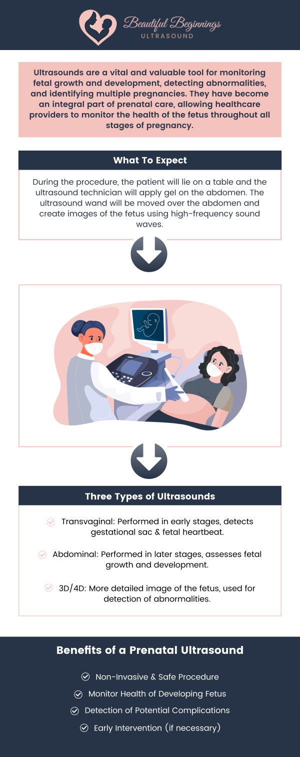
Table of Contents:
What is an ultrasound in pregnancy?
When do you have your first prenatal ultrasound?
What can be detected in a pregnancy ultrasound?
How many ultrasounds do you have during your pregnancy?
An ultrasound (also called a sonogram) is a prenatal test offered to most pregnant women. It uses sound waves to show an image of your baby in the womb. Using ultrasound, your doctor can check your baby’s health and development.
The timing of your first prenatal ultrasound can vary depending on your pregnancy, medical history, and the recommendation of your healthcare professional. Many people have an early ultrasound during the first trimester to confirm the pregnancy, estimate gestational age, check the location of the pregnancy, and evaluate early development. The exact timing may also depend on when your last menstrual period occurred and whether you are experiencing any symptoms that require earlier medical evaluation.
At Beautiful Beginnings, we offer abdominal ultrasounds starting as early as seven weeks for pregnancy verification. During the scan, our experienced sonographers use noninvasive ultrasound technology to capture images of the developing pregnancy and look for early milestones, including fetal cardiac activity when visible. If it is still too early to see certain structures clearly, a follow-up scan may provide additional information as the pregnancy progresses.
Ultrasound uses high-frequency sound waves to create images of the developing fetus. These sound waves are emitted by the ultrasound machine and bounce off the fetus, creating an image on the screen. This technology allows healthcare providers to monitor fetal growth, determine gestational age, and detect any abnormalities in the fetus. For example, ultrasound can detect structural abnormalities such as cleft lip or heart defects. It can also detect potential complications such as ectopic pregnancy or miscarriage.
One of the main benefits of ultrasound is that it is non-invasive and does not harm the fetus or mother. Ultrasound is also an effective tool for identifying potential pregnancy complications, allowing healthcare providers to intervene early and provide appropriate care. For example, if a healthcare provider identifies a potential complication such as preterm labor, they can take steps to prevent premature birth. Ultrasound can also help identify birth defects, allowing parents and healthcare providers to prepare for any necessary medical interventions.
Prenatal care is an essential aspect of a healthy pregnancy. One of the most common procedures during prenatal care is an ultrasound. Ultrasounds are used to monitor fetal growth and development, detect abnormalities, and identify multiple pregnancies.
Ultrasound during pregnancy is a noninvasive imaging procedure that uses high-frequency sound waves to create images of the developing baby. At Beautiful Beginnings, we offer abdominal ultrasounds starting as early as seven weeks for pregnancy verification. These early scans can help visualize the gestational sac, estimate gestational age, and identify fetal cardiac activity when visible. As pregnancy progresses, abdominal ultrasound can provide clearer views of the baby’s growth, position, and development.
We also offer 3D/4D ultrasounds later in pregnancy, which provide more detailed views of your baby’s facial features, movements, and overall appearance. Ultrasound timing can vary depending on the stage of pregnancy and the purpose of the scan, with additional ultrasounds scheduled as appropriate throughout the pregnancy.
Ultrasound during pregnancy has several benefits. It can detect fetal abnormalities, such as neural tube defects and Down syndrome. It can also assess fetal growth and development, including the size of the fetus, the amount of amniotic fluid, and the position of the placenta. Ultrasound can also identify multiple pregnancies, which can lead to a higher risk of complications during pregnancy and delivery. However, there are also risks associated with ultrasound during pregnancy. Exposure to high-frequency sound waves has not been proven to cause harm to the fetus, but some studies suggest that it may affect fetal development. Additionally, there is a risk of misinterpretation of results, which can lead to unnecessary interventions or missed abnormalities.
Preparing for an ultrasound during pregnancy is simple. It is recommended to drink water before the procedure to fill the bladder, which helps to create a clearer image of the fetus. Loose clothing is also recommended to allow easy access to the abdomen. During the procedure, the patient will lie on a table and the ultrasound technician will apply gel to the abdomen. The ultrasound wand will then be moved over the abdomen to create images of the fetus. The technician will identify fetal organs and structures, measure fetal size, and detect abnormalities if present. The results of the ultrasound will be interpreted by a radiologist or obstetrician and shared with the patient during a follow-up appointment.
Ultrasound during pregnancy is a valuable tool for monitoring fetal growth and development, detecting abnormalities, and identifying multiple pregnancies. While there are risks associated with ultrasound, they are generally considered to be minimal.
Preparing for an ultrasound is simple and the procedure itself is non-invasive. By providing a comprehensive guide to ultrasounds during pregnancy, we hope to alleviate any concerns or confusion that expectant mothers may have about this important aspect of prenatal care. We serve patients from Shrewsbury NJ, Red Bank NJ, Little Silver NJ, Eatontown NJ, Middletown NJ, Colts Neck NJ, Rumson NJ, Holmdel NJ, Long Branch NJ, West Long Branch NJ, Ocean City NJ, Oceanport NJ, and Surrounding Areas.

Check Out Our 5 Star Reviews


Additional Services You May Need
▸ 3D Ultrasound Imaging
▸ 4D Ultrasound Imaging
▸ 5D Ultrasound Imaging
▸ 3D and 4D Ultrasound
▸ Elective Ultrasound
▸ Early Gender Determination
▸ Prenatal Ultrasound
▸ Ultrasound Imaging
▸ Ultrasound Scans
▸ 12k Imaging
▸ Sonogram

Additional Services You May Need
▸ 3D Ultrasound Imaging
▸ 4D Ultrasound Imaging
▸ 5D Ultrasound Imaging
▸ 3D and 4D Ultrasound
▸ Elective Ultrasound
▸ Early Gender Determination
▸ Prenatal Ultrasound
▸ Ultrasound Imaging
▸ Ultrasound Scans
▸ 12k Imaging
▸ Sonogram



 Send Text
 How May I Help You?